
What it is
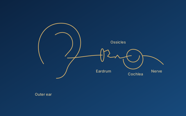
Cholesteatoma is a pocket of skin — keratinising squamous epithelium — trapped inside the middle ear or mastoid. Skin sheds constantly; in the ear canal the debris falls out, but inside a closed pocket it accumulates, and the growing mass presses on and slowly dissolves the surrounding bone. The ossicles, the wall of the inner ear, the facial nerve canal and the thin bone separating the ear from the brain are all within reach.
It is not a tumour and it does not spread through the body. Its danger is local and mechanical, and it does not stop on its own.
How it develops
Most cholesteatomas begin as a retraction pocket: a weakened part of the eardrum, usually at the top, is sucked inwards by a poorly ventilated middle ear until it becomes a sac that can no longer clean itself. Some arise at the edge of a perforation, some are congenital (present behind an intact drum in a child with no history of infection), and some follow surgery or trauma that implants skin in the wrong place.
Symptoms
A smelly, persistent or recurring discharge that does not respond well to drops is the classic sign. Hearing loss follows as the ossicles are eroded. Dizziness, facial weakness, severe pain or headache suggest a complication and need urgent assessment.
Diagnosis
The pocket and its debris are usually visible on examination with a microscope or endoscope. A CT scan shows the extent of the disease and the bone that has been lost, and warns the surgeon about a low-lying facial nerve, an eroded semicircular canal or a thin skull base. Hearing tests document the starting point.
Why surgery, and what it aims to do
Cholesteatoma cannot be cured by medication or by a hearing aid. Surgery removes the skin sac and creates an ear that will not re-form it. The primary objective is a safe, dry, self-cleaning ear — disease control. Hearing improvement is a second, separate objective: sometimes achieved at the same operation by reconstructing the ossicles, sometimes deferred to a planned second stage once the ear is known to be clear, and sometimes not achievable, in which case a hearing device becomes part of the plan.
Being clear about this distinction protects against disappointment. A technically excellent operation that leaves a safe ear with unchanged hearing has met its first goal.
Surgical strategies
Canal-wall-up mastoidectomy preserves normal anatomy and ear care but carries a higher rate of residual and recurrent disease, historically managed with a planned second look and now increasingly with DWI-MRI surveillance. Canal-wall-down creates an open cavity with lower recurrence but lifelong cavity care and water restrictions unless obliterated. Mastoid obliteration with bone pâté or cartilage after either approach reduces recurrence and cavity problems in several large series. Transcanal endoscopic techniques improve visualisation of the sinus tympani and facial recess and are used alone for limited disease or combined with microscopic mastoid surgery. The choice depends on extent, Eustachian-tube function, the contralateral ear, age and the likelihood of reliable follow-up.
After surgery
Follow-up is not optional. Residual disease can regrow silently behind a healed drum for years. Examination at intervals, and MRI where the ear cannot be fully inspected, are part of the treatment rather than an afterthought.
A note on who operates
This page is educational. Cholesteatoma surgery is performed by otologists in many centres; where it is discussed on this site it does not imply that a particular procedure is personally offered. The second opinion service can help review a diagnosis, a proposed operation or a disappointing result, and point towards appropriate referral. Cholesteatoma and chronic otitis as seen in the Colombiers practice are described at grolman-orl.fr.
This page provides general educational information. It cannot replace an individual assessment, which depends on a full history, examination, audiometry and, where relevant, imaging. If you have sudden hearing loss, severe pain, facial weakness, severe dizziness or discharge with fever, seek medical care promptly.